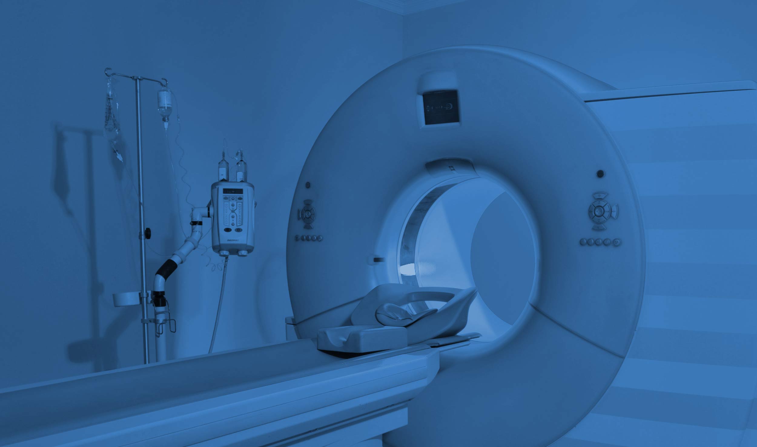
Cross-sectional image of body tissues using x-ray equipment
CT uses x-ray equipment that rotates around the body to obtain image data at different angles. Processing of this data by a computer results in a cross-sectional image of body tissues. It is not usually a claustrophobic experience and is very quick to perform. The whole examination is likely to take about 15 minutes, although the scan itself lasts only a few seconds.
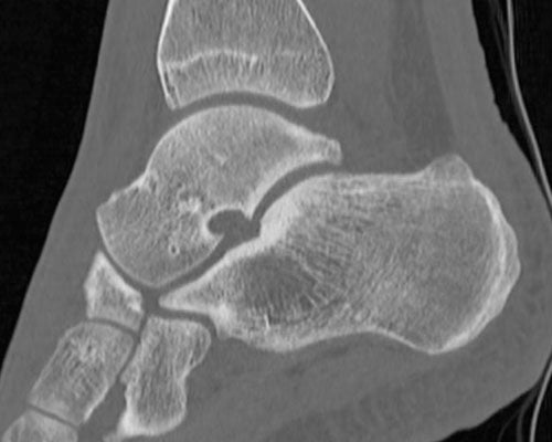
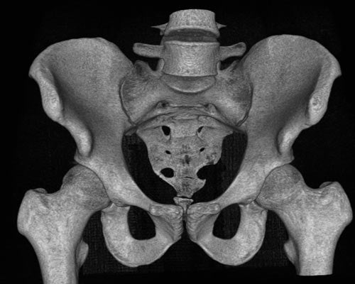
CT demonstrates fine bony detail well and may be used to diagnose and assess bones (eg fractures), joints, the spine and to guide some interventional procedures. It is sometimes useful if there are absolute contraindications to MRI examination or if there has been previous surgery to the area to be scanned with metalwork.
The typical radiation dose for a CT examination is equivalent to the amount of natural background radiation received over a period of one year. You should tell the staff if you are, or think you may be, pregnant as CT is best avoided within the first trimester, unless absolutely essential.
"We offer a wide range of both diagnostic and interventional musculoskeletal services at different sites within the city."